• Bitcoin Falls to 1-Month Low Below $8K

    Fatehlaljeengar
    The price of bitcoin has fallen to a one-month low below $7,900, amid a wider sell-off in the global financial markets.
    As of writing, the price of the world's largest cryptocurrency by market capitalization is changing hands at $7,837, the lowest in the past 30 days and a 10 percent decline on a 24-hour basis, according to CoinDesk's Bitcoin Price Index.
    The price decline comes as the global financial markets suffer a wider sell-off, with Brent crude oil dropping over 30 percent on Sunday, its biggest single-day decline since 1991.
    Underscoring the severity of the global flight from assets perceived as risky is the drop in the 10-year U.S. Treasury yield below 0.5 percent for the first time ever, down over two percentage points from a year ago.
    Bitcoin's sudden price dip also comes as the network's computing power and mining difficulty (a measure of competition among miners) are both expected to reach a new high in just five hours.
    With more processing power chasing a less-valuable asset, mining farms that are using old mining equipment are in for an even tougher time.
    Data from the mining pool Poolin shows that the most widely used mining computers such as the AntMiner S9 and Avalon 851 are all at a critical breakeven point, meaning they are not generating any daily profits at bitcoin's current price, amid all-time-high mining difficulty.

    WHO statement on cases of COVID-19 surpassing 100 000

    Fatehlaljeengar
    As of today’s reports, the global number of confirmed cases of COVID-19 has surpassed 100 000. As we mark this sombre moment, the World Health Organization (WHO) reminds all countries and communities that the spread of this virus can be significantly slowed or even reversed through the implementation of robust containment and control activities. 
    China and other countries are demonstrating that spread of the virus can be slowed and impact reduced through the use of universally applicable actions, such as working across society to identify people who are sick, bringing them to care, following up on contacts, preparing hospitals and clinics to manage a surge in patients, and training health workers.
    WHO calls on all countries to continue efforts that have been effective in limiting the number of cases and slowing the spread of the virus. 
    Every effort to contain the virus and slow the spread saves lives. These efforts give health systems and all of society much needed time to prepare, and researchers more time to identify effective treatments and develop vaccines. 
    Allowing uncontrolled spread should not be a choice of any government, as it will harm not only the citizens of that country but affect other countries as well. 
    We must stop, contain, control, delay and reduce the impact of this virus at every opportunity. Every person has the capacity to contribute, to protect themselves, to protect others, whether in the home, the community, the healthcare system, the workplace or the transport system. 
    Leaders at all levels and in all walks of life must step forward to bring about this commitment across society. 
    WHO will continue to work with all countries, our partners and expert networks to coordinate the international response, develop guidance, distribute supplies, share knowledge and provide people with the information they need to protect themselves and others.

    Constipation

    Fatehlaljeengar
    Definition
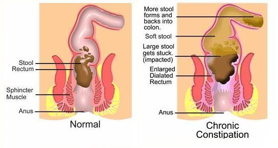
    Constipationcostiveness, or irregularity, is a condition of the digestive system in which a person experiences hard feces that are difficult to expel.Constipation
    • This usually happens because the colon absorbs too much water from the food. If the food moves through the gastro-intestinal tract too slowly, the colon may absorb too much water, resulting in feces that are dry and hard.
    • Defecation may be extremely painful, and in severe cases (fecal impaction) lead to symptoms of bowel obstruction.
    Causes of constipation:
    • may be dietary
    • hormonal
    • anatomical a side effect of medications (e.g. some opiates)
    • or an illness or disorder.
    Clinical Manifestations
    • Fewer than three bowel movements per week, abdominal distention, and pain and pressure
    • Decreased appetite, headache, fatigue, indigestion, sensation of incomplete emptying
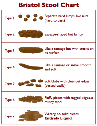
    • Straining at stool; elimination of small volume of hard, dry stool
    • Complications such as hypertension, hemorrhoids and fissures, fecal impaction, and megacolon
    Assessment and Diagnostic Methods
    • Diagnosis is based on history, physical examination, possibly a barium enema or sigmoidoscopy, stool for occult blood, anorectal manometry (pressure studies), defecography, and colonic transit studies.
    • Newer tests such as pelvic floor MRI may identify occult pelvic floor defects.
    stoolclassification
    Medical Management
    • Treatment should target the underlying cause of constipation and aim to prevent recurrence, including education, bowel habit training, increased fiber and fluid intake, and judicious use of laxatives.
    • Discontinue laxative abuse; increase fluid intake; include fiber in diet; try biofeedback, exercise routine to strengthen abdominal muscles.
    • If laxative is necessary, use bulk-forming agents, saline and osmotic agents, lubricants, stimulants, or fecal softeners.
    • Specific medication therapy to increase intrinsic motor function (eg, cholinergics, cholinesterase inhibitors, or prokinetic agents).
    Nursing Management
    Assessment
    Use tact and respect with patient when talking about bowel habits and obtaining health history.
    Note the following:
    • Onset and duration of constipation, current and past elimination patterns, patient’s expectation of normal bowel elimination, and lifestyle information (eg, exercise and activity level, occupation, food and fluid intake, and stress level).
    • Past medical and surgical history, current medications, history of laxative or enema use.
    • Report of any of the following: rectal pressure or fullness, abdominal pain, straining at defecation, and flatulence.
    • Sets specific goals for teaching; goals for the patient include restoring or maintaining a regular pattern of elimination by responding to the urge to defecate, ensuring adequate intake of fluids and high fiber foods, learning about methods to avoid constipation, relieving anxiety about bowel elimination patterns, and avoiding complications.

    Alzheimer Disease

    Fatehlaljeengar

    Description
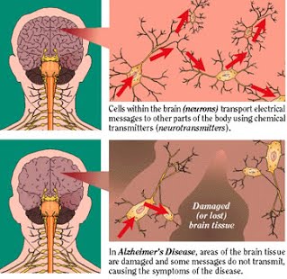
    • Alzheimer’s disease is an irreversible, progressive brain disease that slowly destroys memory and thinking skills, and eventually even the ability to carry out the simplest tasks. In most people with Alzheimer’s, symptoms first appear after age 60.
    • Alzheimer’s disease is the most common cause of dementia among older people. Dementia is the loss of cognitive functioning—thinking, remembering, and reasoning—to such an extent that it interferes with a person’s daily life and activities. Estimates vary, but experts suggest that as many as 5.1 million Americans may have Alzheimer’s.
    • Dementia involves progressive decline in two or more of the following areas of cognition: memory, language, calculation, visual-spatial perception, judgment, abstraction, and behavior.
    Causes
    The cause of AD is unknown but knowledge about the hereditary links is growing. Patients with Down syndrome eventually develop DAT if they live long enough. There is a higher-than-normal concentration of aluminum in the brain of a person with DAT, but the effect is unknown. A distinct protein, AZ 50, has been identified at autopsy in the brains of DAT patients. This protein has been isolated from neurons that were not yet damaged, suggesting that its presence early in the degenerative process might cause the neuronal damage. The life expectancy of a DAT patient is reduced 30% to 60%.
    alzheimers-diseaseCharacteristics/ Signs and Symptoms
    The disease course is divided into four stages, with progressive patterns of cognitive and functional impairments.
    Pre-dementia
    Stage 1 is characterized by recent memory loss, increased irritability, impaired judgment, loss of interest in life, decline of problem-solving ability, and reduction in abstract thinking. Remote memory and neurological exam remain unchanged from baseline.
    Early
    Stage 2 lasts 2 to 4 years and reveals a decline in the patient’s ability to manage personal and business affairs, an inability to remember shapes of objects, continued repetition of a meaningless word or phrase (perseveration), wandering or circular speech patterns (circumlocution dysphasia), wandering at night, restlessness, depression, anxiety, and intensification of cognitive and emotional changes of stage 1.
    Moderate
    Stage 3 is characterized by impaired ability to speak (aphasia), inability to recognize familiar objects (agnosia), inability to use objects properly (apraxia), inattention, distractibility, involuntary emotional outbursts, urinary or fecal incontinence, lint-picking motion, and chewing movements. Progression through stages 2 and 3 varies from 2 to 12 years.
    Advanced
    Stage 4, which lasts approximately 1 year, reveals a patient with a masklike facial expression, no communication, apathy, withdrawal, eventual immobility, assumed fetal position, no appetite, and emaciation.
    Diagnostic Examination
    • Alzheimer’s disease is usually diagnosed clinically from the patient history, collateral history from relatives, and clinical observations, based on the presence of characteristic neurological and neuropsychological features and the absence of alternative conditions.
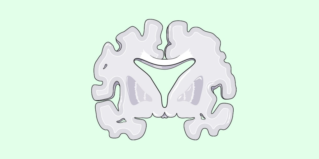
    • Advanced medical imaging with computed tomography (CT) or magnetic resonance imaging (MRI), and with single photon emission computed tomography (SPECT) or positron emission tomography (PET) can be used to help exclude other cerebral pathology or subtypes of dementia.Moreover, it may predict conversion from prodromal stages (mild cognitive impairment) to Alzheimer’s disease.
    • Assessment of intellectual functioning including memory testing can further characterize the state of the disease.Medical organisations have created diagnostic criteria to ease and standardize the diagnostic process for practicing physicians. The diagnosis can be confirmed with very high accuracy post-mortem when brain material is available and can be examined histologically.
    Primary Nursing Diagnosis
    Self-care deficit related to impaired cognitive and motor function
    • Outcomes. Self-care: Activities of daily living—Bathing, Hygiene, Eating, Toileting; Cognitive ability; Comfort level; Role performance; Social interaction skills; Hope
    • Interventions. Self-care assistance: Bathing and Hygiene; Oral health management; Behavior management; Body image enhancement; Emotional support; Mutual goal setting; Exercise therapy; Discharge planning
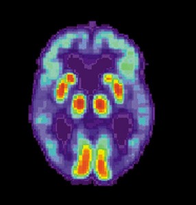
    PET_Alzheimer
    PET scan of the brain of a person with AD showing a loss of function in the temporal lobe
    Other Nursing Diagnosis
    Risk for Injury related to:
    • Unable to recognize / identify hazards in the environment.
    • Disorientation, confusion, impaired decision making.
    • Weakness, the muscles are not coordinated, the presence of seizure activity.
    Medical Management
    There is no cure for Alzheimer’s disease; available treatments offer relatively small symptomatic benefit but remain palliative in nature.
    The initial management of the patient begins with education of the family and caregivers regarding the disease, the prognosis, and changes in lifestyle that are necessary as the disease progresses.
    Basic collaborative principles include:
    • Keep requests for the patient simple
    • Avoid confrontation and requests that might lead to frustration
    • Remain calm and supportive if the patient becomes upset
    • Maintain a consistent environment
    • Provide frequent cues and reminders to reorient the patient
    • Adjust expectations for the patient as he or she declines in capacity
    Pharmacologic Treatment
    • Generally, therapy is focused on symptoms with an attempt to maintain cognition.
    • Donepezil (cholinesterase inhibitor; elevates acetylcholine concentration in cerebral cortex by slowing degradation of acetylcholine released by intact neurons)which improves cognitive symptoms; improves cognitive function in the early stages of the disease only; drug effects diminish as the disease progresses
    • Antidepressants (selective serotonin reuptake inhibitors; increases activity of serotonin in the brain) which treats depression, anxiety, and irritability
    • Other Tests: Supporting tests include computed tomography (CT) scan; magnetic resonance imaging (MRI); positron emission tomography (PET). During the early stages of dementia, CT and MRI may be normal, but in later stages, an MRI may show a decrease in the size of the cerebral cortex or of the area of the brain responsible for memory, particularly the hippocampus. Genetic testing for the ApoE gene is available and the presence of the gene is a risk factor for AD. Genetic tests may be helpful in diagnosis, but further studies are needed to confirm their reliability.
    Nursing Intervention
    1. Establish an effective communication system with the patient and his family to help them adjust to the patient’s altered cognitive abilities.
    2. Provide emotional support to the patient and his family.
    3. Administer ordered medications and note their effects. If the patient has trouble swallowing, crush tablets and open capsules and mix them with a semi soft food.
    4. Protect the patient from injury by providing a safe, structured environment.
    5. Provide rest periods between activities because the patient tires easily.
    6. Encourage the patient to exercise as ordered to help maintain mobility.
    7. Encourage patient independence and allow ample time for him to perform tasks.
    8. Encourage sufficient fluid intake and adequate nutrition.
    9. Take the patient to the bathroom at least every 2 hours and make sure he knows the location of the bathroom.
    10. Assist the patient with hygiene and dressing as necessary.
    11. Frequently check the the patient’s vital signs.
    12. Monitor the patient’s fluid and food intake to detect imbalances.
    13. Inspect the patient’s skin for evidence of trauma, such as bruises or skin breakdown.
    14. Encourage the family to allow the patient as much independence as possible while ensuring safety to the patient and others.

    कोरोना वायरस

    Fatehlaljeengar

    कोरोना  वायरस

    कोरोना वायरस (सीओवी) का संबंध वायरस के ऐसे परिवार से है, जिसके संक्रमण से जुकाम से लेकर सांस लेने में तकलीफ जैसी समस्या हो सकती है. इस वायरस को पहले कभी नहीं देखा गया है. इस वायरस का संक्रमण दिसंबर में चीन के वुहान में शुरू हुआ था. डब्लूएचओ के मुताबिक, बुखार, खांसी, सांस लेने में तकलीफ इसके लक्षण हैं. अब तक इस वायरस को फैलने से रोकने वाला कोई टीका नहीं बना है. 

    बीमारी  के लक्षण

     इसके संक्रमण के फलस्वरूप बुखार, जुकाम, सांस लेने में तकलीफ, नाक बहना और गले में खराश जैसी समस्या उत्पन्न होती हैं. यह वायरस एक व्यक्ति से दूसरे व्यक्ति में फैलता है. इसलिए इसे लेकर बहुत सावधानी बरती जा रही है. यह वायरस दिसंबर में सबसे पहले चीन में पकड़ में आया था. इसके दूसरे देशों में पहुंच जाने की आशंका जताई जा रही है. 

    बचाव  के उपाय

    स्‍वास्‍थ्‍य मंत्रालय ने कोरोना वायरस से बचने के लिए दिशानिर्देश जारी किए हैं. इनके मुताबिक, 

         हाथों को साबुन से धोना चाहिए. 

         अल्‍कोहल आधारित हैंड रब का इस्‍तेमाल भी किया जा सकता है. 

         खांसते और छीकते समय नाक और मुंह रूमाल या टिश्‍यू पेपर से ढककर रखें. जिन व्‍यक्तियों में कोल्‍ड और फ्लू के लक्षण हों उनसे दूरी बनाकर रखें.

         अंडे और मांस के सेवन से बचें. जंगली जानवरों के संपर्क में आने से बचें.

    Appendicitis

    Fatehlaljeengar
    Description
    • Appendicitis is inflammation of the appendix.
    • When the appendix becomes inflamed or infected, rupture may occur within a matter of hours, leading to peritonitis and sepsis.
    Risk Factors
    • Obstruction by fecalith or foreign bodies, bacteria or toxins.
    • Low-fiber diet
    • High intake of refined carbohydrates
    Signs and Symptoms/ Assessment 
    1. Pain in the periumbilical area that descends to the right lower quadrant.
    2. Abdominal pain that is most intense at McBurney’s point
    3. Rebound tenderness and abdominal rigidity
    4. Low-grade fever
    5. Elevated white blood cell count
    6. Anorexia, nausea, and vomiting
    7. Client in side-lying position, with abdominal guarding and legs flexed
    8. Constipation or diarrhea
    Diagnostic Evaluation
    • Diagnosis is based on a complete physical examination and laboratory and radiologic tests.
    • Leukocyte count greater than 10,000/mm 3, neutrophil count greater than 75%; abdominal radiographs, ultrasound studies, and CT scans may reveal right lower quadrant density or localized distention of the bowel.
    Primary Nursing Diagnosis
    Primary Preoperative Nursing Diagnosis
    • Pain (acute) related to inflammation
    Primary Postoperative Nursing Diagnosis
    • Risk for infection related to the surgical incision
    Other Diagnoses that may occur in Nursing Care Plans For Appendicitis
    • Imbalanced nutrition: Less than body requirements
    • Impaired skin integrity
    • Ineffective tissue perfusion: GI
    • Risk for deficient fluid volume
    • Risk for injury
    Medical Management
    An appendectomy (surgical removal of the appendix) is the preferred method of management for acute appendicitis if the inflammation is localized. An open appendectomy is completed with a transverse right lower quadrant incision, usually at the McBurney point. A laparoscopic appendectomy may be used in females of childbearing age, those in whom the diagnosis is in question, and for obese patients. If the appendix has ruptured and there is evidence of peritonitis or an abscess, conservative treatment consisting of antibiotics and intravenous (IV) fluids is given 6 to 8 hours prior to an appendectomy. Generally, an appendectomy is performed within 24 to 48 hours after the onset of symptoms under either general or spinal anesthesia. Preoperative management includes IV hydration, antipyretics, antibiotics, and, after definitive diagnosis, analgesics.

    Complications of Appendectomy
    • The major complication is perforation of the appendix, which can lead to peritonitis or an abscess.
    • Perforation generally occurs 24 hours after onset of pain, symptoms include fever (37.7°C [100° F] or greater), toxic appearance, and continued pain and tenderness.
    Pharmacologic Intervention
    • Crystalloid intravenous fluids an isotonic solutions such as normal saline solution or lactated Ringer’s solution 100–500 mL/hr of IV, depending on volume state of the patient, is used to replaces fluids and electrolytes lost through fever and vomiting; replacement continues until urine output is 1 cc/kg of body weight and electrolytes are replaced
    • Antibiotics (broad-spectrum antibiotic coverage) to control local and systemic infection and reduces the incidence of postoperative wound infection
    • Other Drugs: Analgesics.
    Nursing Intervention
    Preoperative interventions
    1. Maintain NPO status.
    2. Administer fluids intravenously to prevent dehydration.
    3. Monitor for changes in level of pain.
    4. Monitor for signs of ruptured appendix and peritonitis
    5. Position right-side lying or low to semi fowler position to promote comfort.
    6. Monitor bowel sounds.
    7. Apply ice packs to abdomen every hour for 20-30 minutes as prescribed.
    8. Administer antibiotics as prescribed
    9. Avoid the application of heat in the abdomen.
    10. Avoid laxatives or enema.
    Postoperative interventions
    1. Monitor temperature for signs of infection.
    2. Assess incision for signs of infection such as redness, swelling and pain.
    3. Maintain NPO status until bowel function has returned.
    4. Advance diet gradually or as tolerated or as prescribed when bowel sound return.
    5. If ruptured of appendix occurred, expect a Penros drain to be inserted, or the incision maybe left to heal inside out.
    6. Expect that drainage from the Penros drain maybe profuse for the first 2 hours.
    Documentation Guidelines
    • Location, intensity, frequency, and duration of pain
    • Response to pain medication, ice applications, and position changes
    • Patient’s ability to ambulate and tolerate food
    • Appearance of abdominal incision (color, temperature, intactness, drainage)
    Discharge and Home Healthcare Guidelines
    • MEDICATIONS. Be sure the patient understands any pain medication prescribed, including doses, route, action, and side effects. Make certain the patient understands that he or she should avoid operating a motor vehicle or heavy machinery while taking such medication.
    • INCISION. Sutures are generally removed in the physician’s office in 5 to 7 days. Explain the need to keep the surgical wound clean and dry. Teach the patient to observe the wound and report to the physician any increased swelling, redness, drainage, odor, or separation of the wound edges. Also instruct the patient to notify the doctor if a fever develops. The patient needs to know these may be symptoms of wound infection. Explain that the patient should avoid heavy lifting and should question the physician about when lifting can be resumed.
    • COMPLICATIONS. Instruct the patient that a possible complication of appendicitis is peritonitis. Discuss with the patient symptoms that indicate peritonitis, including sharp abdominal pains, fever, nausea and vomiting, and increased pulse and respiration. The patient must know to seek medical attention immediately should these symptoms occur.
    • NUTRITION. Instruct the patient that diet can be advanced to her or his normal food pattern as long as no gastrointestinal distress is experienced.

    Rubella

    Fatehlaljeengar

    Rubella

    Key facts

    • Rubella is a contagious viral infection that occurs most often in children and young adults.
    • Rubella is the leading vaccine-preventable cause of birth defects. Rubella infection in pregnant women may cause fetal death or congenital defects known as congenital rubella syndrome.
    • There is no specific treatment for rubella but the disease is preventable by vaccination.

    • Rubella is an acute, contagious viral infection. While rubella virus infection usually causes a mild fever and rash in children and adults, infection during pregnancy, especially during the first trimester, can result in miscarriage, fetal death, stillbirth, or infants with congenital malformations, known as congenital rubella syndrome (CRS).
      The rubella virus is transmitted by airborne droplets when infected people sneeze or cough. Humans are the only known host.
    • Symptoms

      In children, the disease is usually mild, with symptoms including a rash, low fever (<39°C), nausea and mild conjunctivitis. The rash, which occurs in 50–80% of cases, usually starts on the face and neck before progressing down the body, and lasts 1–3 days. Swollen lymph glands behind the ears and in the neck are the most characteristic clinical feature. Infected adults, more commonly women, may develop arthritis and painful joints that usually last from 3–10 days.
      Once a person is infected, the virus spreads throughout the body in about 5-7 days. Symptoms usually appear 2 to 3 weeks after exposure. The most infectious period is usually 1–5 days after the appearance of the rash.
      When a woman is infected with the rubella virus early in pregnancy, she has a 90% chance of passing the virus on to her fetus. This can cause the death of the fetus, or it may cause CRS. Infants with CRS may excrete the virus for a year or more.
    • Congenital rubella syndrome

      Children with CRS can suffer hearing impairments, eye and heart defects and other lifelong disabilities, including autism, diabetes mellitus and thyroid dysfunction – many of which require costly therapy, surgeries and other expensive care.
      The highest risk of CRS is in countries where women of childbearing age do not have immunity to the disease (either through vaccination or from having had rubella). Before the introduction of the vaccine, up to 4 babies in every 1000 live births were born with CRS.

      Vaccination

      The rubella vaccine is a live attenuated strain, and a single dose gives more than 95% long-lasting immunity, which is similar to that induced by natural infection.
      Rubella vaccines are available either in monovalent formulation (a vaccine directed at only one pathogen) or more commonly in combinations with other vaccines such as with vaccines against measles (MR), measles and mumps (MMR), or measles, mumps and varicella (MMRV).
      Adverse reactions following vaccination are generally mild. They may include pain and redness at the injection site, low-grade fever, rash and muscle aches. Mass immunization campaigns in the Region of the Americas involving more than 250 million adolescents and adults did not identify any serious adverse reactions associated with the vaccine.
    • WHO response

      WHO recommends that all countries that have not yet introduced rubella vaccine should consider doing so using existing, well-established measles immunization programmes. To-date, four WHO regions have established goals to eliminate this preventable cause of birth defects. In 2015, the WHO Region of the Americas became the first in the world to be declared free of endemic transmission of rubella.
      The number of countries using rubella vaccines in their national programme continues to steadily increase. As of December 2018, 168 out of 194 countries had introduced rubella vaccines and global coverage was estimated at 69%. Reported rubella cases declined 97%, from 670 894 cases in 102 countries in 2000 to 14 621 cases in 151 countries in 2018. CRS rates are highest in the WHO African and South-East Asian regions where vaccination coverage is lowest.
      In April 2012, the Measles Initiative – now known as the Measles & Rubella Initiative – launched a Global Measles and Rubella Strategic Plan which covers the period 2012-2020. The Plan includes a series of global goals for 2020.
    • By the end of 2020

      • Achieve measles and rubella elimination in at least 5 WHO regions.
      Based on the 2018 Global Vaccine Action Plan (GVAP) Assessment Report by the WHO Strategic Advisory Group of Experts (SAGE) on Immunization, rubella control is lagging, with 26 countries still do introduce the vaccine, while two regions (African and Eastern Mediterranean) have not yet set rubella elimination or control targets.
      SAGE recommends that rubella vaccination should be incorporated into immunization programmes, as quickly as possible, to ensure additional gains in controlling rubella can be made. As one of the founding members of the Measles & Rubella Initiative, WHO provides technical support to governments and communities to improve routine immunization programmes and hold targeted vaccination campaigns. In addition, the WHO Global Measles and Rubella Laboratory Network supports the diagnosis of rubella and CRS cases and tracking of the spread of rubella viruses.